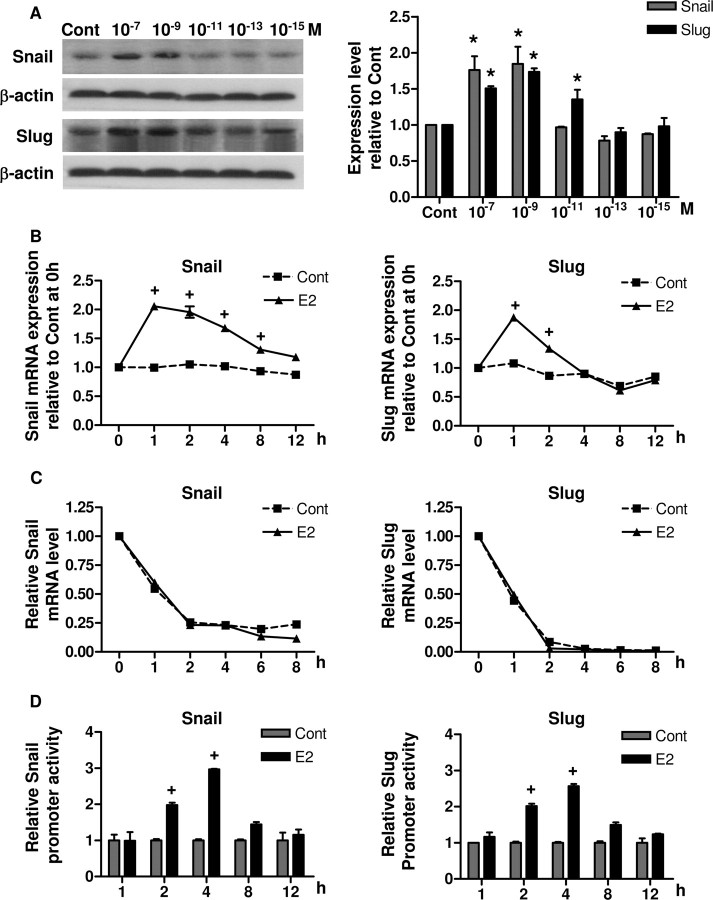
Fig. 4.
E2 Regulates Expression and Gene Promoter Activities of Snail and Slug
A, BG-1 cells were treated with vehicle (Cont) or increasing concentrations of E2 (10−15 to 10−7 m) for 8 h. Whole-cell lysates were extracted, and Snail and Slug were detected by Western blot. β-Actin was used as a loading control. The immunoblot shown is representative of three independent experiments. Cumulative results for quantitative densitometry of the three experiments are shown in left panel. B, BG-1 cells were cultured in the presence of vehicle (Cont) or 10−7 m E2 for the various time periods indicated and harvested for RNA extraction. C, Cells were treated with 10−7 m E2 or vehicle (Cont) for 1 h. Then, at time 0 h, the transcription inhibitor actinomycin D (5 μg/ml) was added to the medium and cells were harvested at the indicated time points. Total RNA was isolated and used for real-time PCR analysis of Snail or Slug mRNA expression with gene-specific primers. GAPDH primers were used to normalize data, and results are expressed as fold change relative to time zero. D, BG-1 cells were transfected with either human Snail or Slug promoter-reporter gene construct and pSV-βGal for normalization. The luciferase activities were determined in cell lysates 2–8 h after initiation of E2 exposure. The luciferase activities were calculated relative to the promoterless vector (pGL3-Basic) and expressed as fold change relative to vehicle control at corresponding time point. The data are shown as mean ± sd of three repeated experiments. *, P < 0.05 compared with control. +, P < 0.05 compared with the corresponding time-matched vehicle control. Cont, Control.

