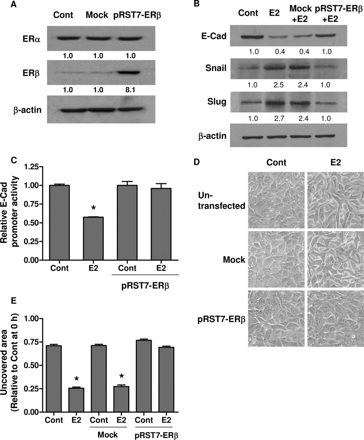
Fig. 8.
ERβ Opposes ERα-Induced EMT
A, BG-1 cells were transfected with pRST7-ERβ or without plasmid DNA (mock). Cell lysates were extracted for ERβ detection by Western blot. B, BG-1 cells were transfected for 8 h and subsequently treated with 10−7 m E2 or vehicle (Cont) for an additional 48 h. E-cadherin (E-Cad), Snail, Slug, and β-actin were detected by Western blot. C, Cells were transfected with pRST7-ERβ, E-cadherin promoter construct, and pSV-βGal. Eight hours after transfection, cells were incubated with vehicle (Cont) or 10−7 m E2 for an additional 48 h. The luciferase activities were calculated relative to the promoterless vector (pGL3-Basic) and expressed as fold change relative to vehicle control. D, Morphological changes of BG-1 cells transfected with pRST7-ERβ or without plasmid DNA (mock) were also observed by phase-contrast microscope, and representative photographs are shown. Original magnification, ×200. E, Scratch was made by scraping the monolayer 24 h after transfection, and cells were incubated with medium containing vehicle (Cont) or 10−7 m E2. Wound closure was photographed after 24 h. The amount of wound repair was expressed as uncovered area compared with initial uncovered area of vehicle-treated control at time zero. Values are the mean ± sd of three separate experiments. *, P < 0.05 compared with control. Numerical values below each lane of the immunoblots represent quantification of the relative protein level by densitometry (normalized to β-actin protein level). Cont, Control.

