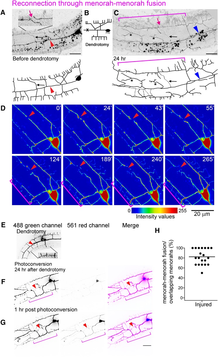
Figure 4.
Menorah–menorah fusion bypasses the injury site and ensures dendrite continuity. (A–C) Dendrotomy of an L4 animal resulted in menorah–menorah fusion. Bar, 10 μm. (A) Just prior to the injury, separated menorahs can be seen (magenta arrow in inset). Red arrowhead points to the lesion site. (B) Schematic showing the site of injury (marked with an X). (C) Formation of giant menorahs (magenta bracket) and ectopic branching (asterisks) within 24 hr. Magenta arrow points to the site of loss of self-avoidance. Blue arrowhead points to the site of primary branch fusion. Pruning of secondary branches also occurs [black arrow, dotted lines, also marked in (A)]. (D) Intensity values view of images from a time-lapse movie of injured L4 wild-type worm (File S4). Time after injury is shown at the upper right corner in minutes. Red arrowheads point to injury site and pruning of branches. Brackets mark two menorahs from distal and proximal ends bypassing the break and contacting one another. (E–G) PVD menorah–menorah fusion confirmed by Kaede photoconversion. Bar, 10 μm. (H) The fraction of menorah–menorah fusion out of the number of loss of self-avoidance events (shown as percentage; horizontal line is the average). n = 20.

