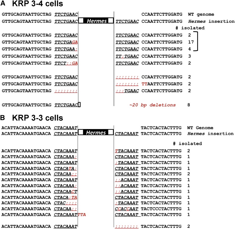
Figure 3.
Hermes excision footprint sequences indicate repair by NHEJ. Hermes excision events were monitored by cloning and sequencing the first-round PCR products from individual colonies in which transposon excision was induced. Cells bearing one of two independent transposon insertions were analyzed: KRP 3-4 cells (A) and KRP 3-3 cells (B). The PCR products for the KRP 3-4 cells are those shown in Figure 2B. The underlined sequence is the 8-bp duplication generated during transposon insertion. Base changes are indicated in red, and deletions are indicated by a colon. Most events show small deletions (0–5 nucleotides) and mutations that implicate NHEJ (Daley et al. 2005; McVey and Lee 2008). A bracket indicates the sequences used to model the mechanism of repair (Figure S4 in File S1). NHEJ, nonhomologous end-joining.

