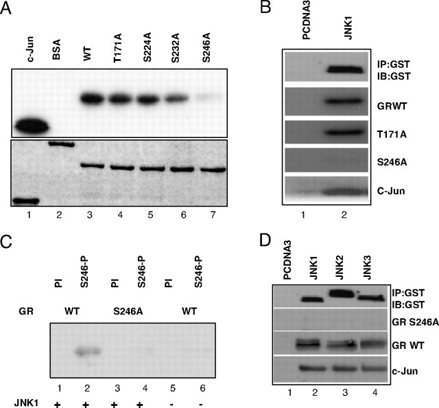
Fig. 1.
GR Is Phosphorylated by the JNK Family of Proteins
A, GST-GR fusion protein carrying WT AF-1 domain of GR (lane 3) or T171A (lane 4), S224A (lane 5), S232A (lane 6), and S246A (lane 7) were expressed and purified from Escherichia coli, and resolved on SDS-PAGE and Coomassie blue stained (lower panel). Indicated proteins were used in in vitro kinase assays with radiolabeled ATP as substrates for JNK1α1 kinase (Upstate Biotechnology), together with GST-c-Jun fusion protein (lane 1) that served as positive control and BSA (lane 2) that served as negative control (top panel). B, COS-7 cells were transfected with control vector pcDNA3 (lane 1) or PEBG JNK1α1 (lane 2), together with pcDNA3HA-MLK3 plasmids. Top panel shows Western blot analysis of JNK1α1 kinase immunoprecipated with GST-specific antibody. GST GR AF-1 WT, T171A, and S246A (middle panels), or c-Jun (lower panel) purified proteins were phosphorylated in vitro with immunoprecipitated kinases in the presence of radiolabeled ATP. Proteins were resolved on SDS-PAGE and exposed to film. C, GST-GR AF-1 WT (lanes 1, 2, 5, and 6) and S246A fusion proteins (lanes 3 and 4) were used as substrates in in vitro kinase assays with nonradiolabeled ATP and JNK1α1 kinase (Upstate Biotechnology). GR was detected by Western blot analysis using preimmune serum (PI) or antibody raised against phosphorylated S246 (S246-P). D, COS-7 cells were transfected with control vector pcDNA3 (lane 1) or PEBG JNK1α1 (lane 2), JNK2α2 (lane 3), and JNK3α1 (lane 4), together with pcDNA3HA-MLK3 plasmids (lanes 2–4). Top panel shows Western blot analysis of JNK1α1 kinase immunoprecipated with GST-specific antibody. GST GR AF-1 S246A (second panel), GST GR AF-1 WT (third panel), or c-Jun (lower panel) purified proteins were phosphorylated in vitro with immunoprecipitated kinases, and Western blot was developed using S246-P antibody (middle panels) and c-Jun phosphospecific antibody (lower panel). IB, Immunoblot; IP, immunoprecipitation.

