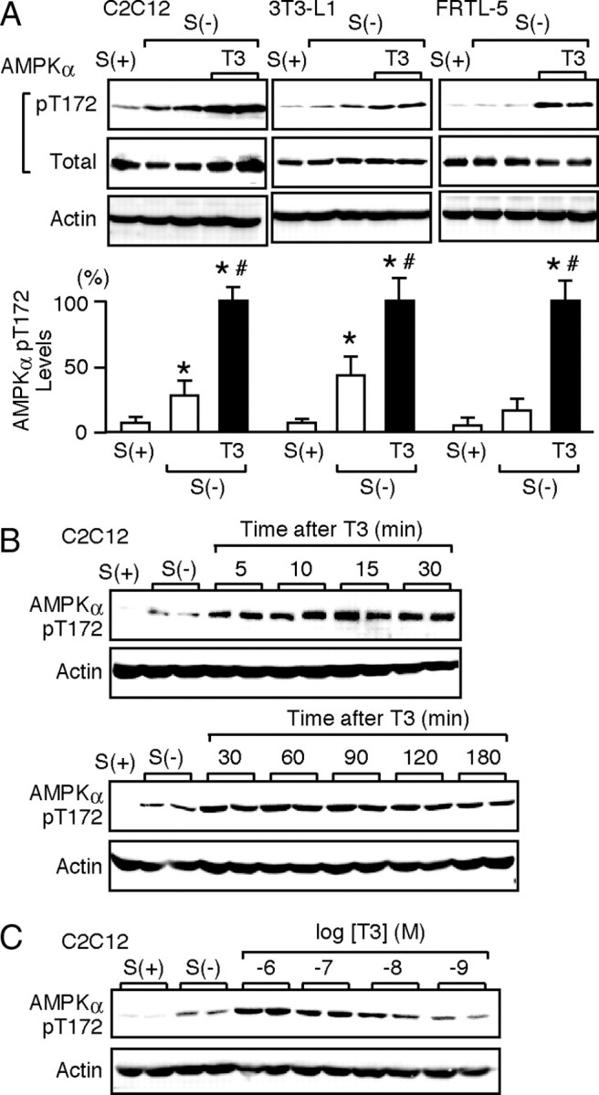
Fig. 1.

T3 Induces the Phosphorylation of Thr-172 in the α-Subunit of AMPK
Differentiated C2C12, 3T3-L1, and FRTL-5 cells were cultured with serum [S(+)] in serum-deprived media for 12 h [S(−)]. A, The cells were treated with 10 nm T3 for 30 min. Whole-cell lysates were subjected to Western blot analysis using antibodies directed against phospho-AMPK α-subunit (pT172), AMPK α-subunit (total), and actin. Most experiments were performed in duplicate cultures. Similar results were obtained from separate experiments. In densitometric analysis, the phospho-AMPK levels were normalized to the actin levels and expressed as percentages of the levels in T3-treated cells. Results are shown as means ± sd (n = 4). *, P < 0.05 vs. the levels in S(+) cells; #, P < 0.05 vs. the levels in S(−) cells. B, C2C12 cells were treated with 10 nm T3 for the indicated times, and Western blot analysis was performed. C, C2C12 cells were treated with 1 nm to 1 μm T3 for 30 min, and Western blot analysis was performed.
