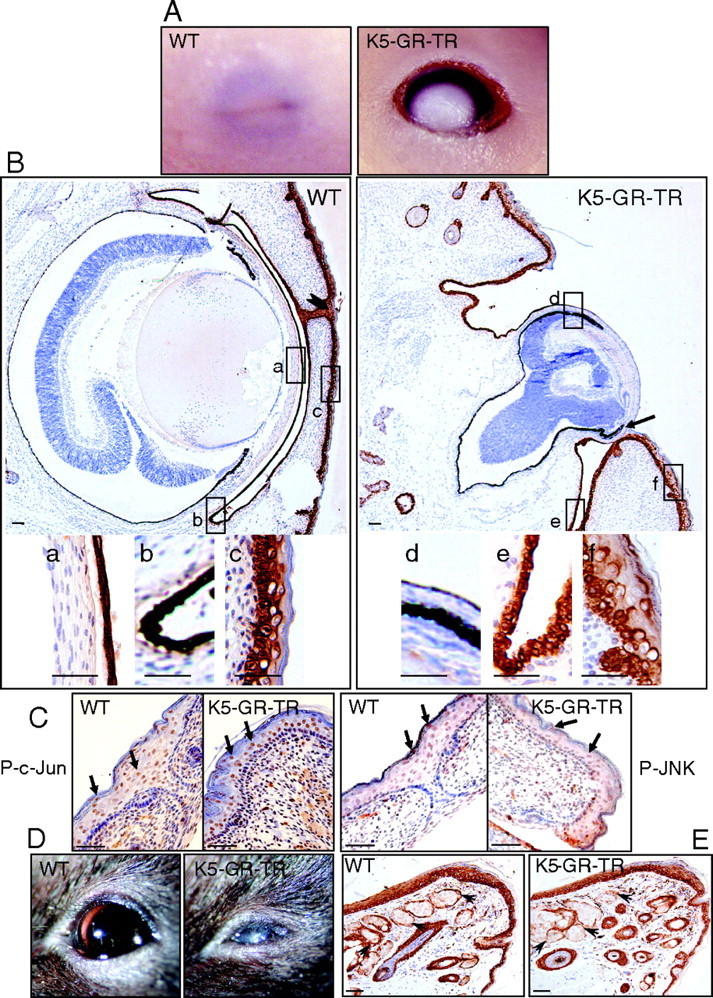
Fig. 2.

Lack of Eyelid Closure in K5-GR-TR Embryos
A, Eyelid fusion failed in 18.5-dpc K5-GR-TR HM embryos as compared with WT mice. B, Immunostaining using K5 in WT and K5-GR-TR embryos demonstrating the epithelia to which transgene expression is targeted: cornea (a and d), conjunctiva (b and e), and eyelid epithelium (c and f). Fused eyelids in WT embryos are pointed to by an arrowhead. a–f are also shown at higher magnification in the lower panel. Note that K5 staining was lacking in the cornea epithelium, whereas it appeared as more restricted in K5-GR-TR eye embryos. Additional ocular defects, including microphthalmia, dysplastic retinal tissue, and attachment of eyelid epithelium to the posterior side of the cornea were observed in K5-GR-TR transgenic embryos (arrow). C, Immunostaining using P-c-Jun and P-JNK antibodies revealed no differences in the expression of these phosphorylated proteins in the eyelid epithelium of K5-GR-TR vs. WT embryos. Arrows indicate nuclear localization of P-c-Jun and P-JNK. D, Adult transgenic mice exhibited corneal opacity. E, The secretory Meibomian glands of the eyelids were unaffected in the transgenics (arrowheads). Scale bar, 50 μm.
