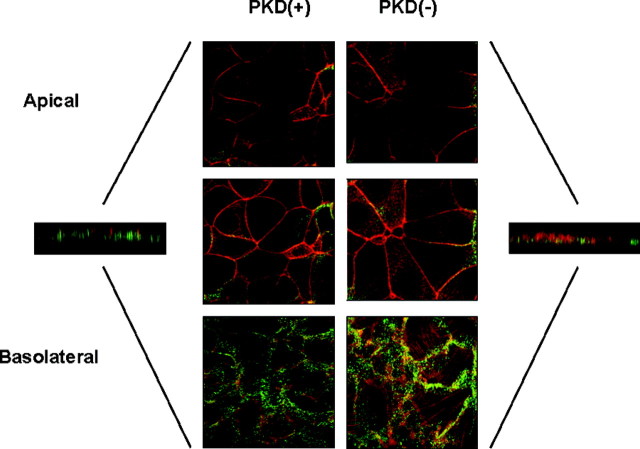
Fig. 6.
Polarization of M1-CCD Cells on Glass
Wild-type M1-CCD cells (A) and M1-CCD cells harboring a plasmid expressing PKD1-specific siRNA (B) were grown on a glass surface until a confluent monolayer was established. The polymerized actin structure of the cells was stained using TRITC-labeled phalloidin (red), and the distribution of the Na+/K+ ATPase pump within the cells was determined by confocal immunofluorescence using a specific monoclonal antibody and detected using an antimouse Alexa 488 conjugate (green). The pump was located only at the basolateral surface of both cell lines.

