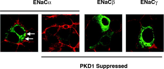
Fig. 8.
Suppression of PKD1 Expression Blocks ENaCα Redistribution
Wild-type M1-CCD cells and cells stably suppressed in their PKD1 expression were transfected with a plasmid expressing ENaCα as an eCFP fusion protein (green). The subcellular distribution of ENaCα was examined by confocal microscopy in untreated cell monolayers and cells treated with aldosterone (10 nm) for 2 min. Cells were counterstained with TRITC-labeled phalloidin to detect polymerized actin (red), and single (XY) focal plane images through the center of the cells are shown. Small dense accumulations of ENaCα were observed within 2 min of aldosterone treatment in wild-type (white arrows) but not in PKD1-suppressed cells.

