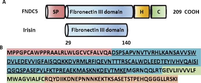
Fig.1.
Structure of the murine FNDC% and irisin protein. (A) Scheme of the murine FNDC5 protein structure (top) and murine irisin protein structure (bottom). SP = signal peptide, H = hydrophobic domain, C = cytoplasmic domain. (B) Murine FNDC5 amino acid sequence with corresponding domains colored. The irisin sequence is underlined.

