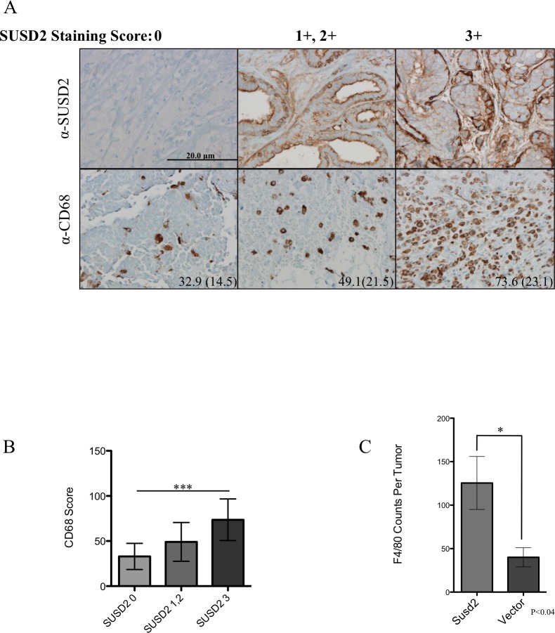
Fig 1. SUSD2 is associated with increased macrophages in breast tumors.
(A) Tissue sections from women undergoing surgical treatment for BCa were analyzed by immunohistochemical analysis using anti-SUSD2 and anti-CD68 (macrophage marker) antibodies. Positive staining is indicated by the brown color. Cells were counterstained blue with hematoxylin. CD68 scores were obtained by counting the number of positive staining cells per high power field (hpf) in three hot spots throughout the tumor. Representative images of each sample are shown. Left, middle and right columns are sections from tumors with a SUSD2-staining score of 0, 1+/2+, and 3+, respectively. The average score with standard deviation in parentheses is located in the bottom right corner of the image for CD68 staining. Images were taken at 200× magnification. (B) Association between CD68 score and SUSD2 staining in patient tumors. Data are represented as mean. Error bars indicate standard deviation (*** p<0.0001). (C) The total number of macrophages per tumor section of 66CL4 mouse tumors stained with anti-F4/80 activated macrophage marker.

