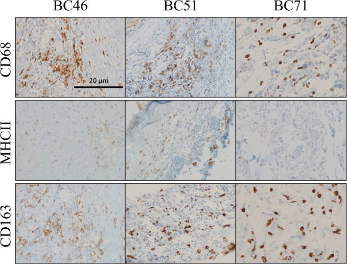
Fig 2. Immunohistochemical analysis of M1 and M2 macrophage polarization in breast tumors.
Tumor sections with strong (3+) SUSD2 staining were analyzed from ten breast cancer patients (representative tumors BC46, BC51, and BC71 are shown). The patient tumors were immunostained using anti-MHCII (M1 marker) and anti-CD163 (M2 marker) antibodies. The brown color indicates positive staining, and the cells were counterstained blue with hematoxylin. Images were taken at 200× magnification. Representative images are shown. Similar areas of each of the tumors were imaged in all three serial sections.

