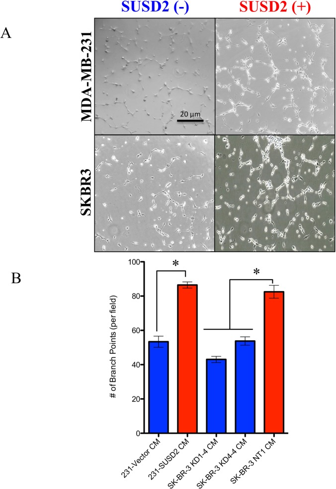
Fig 6. Conditioned media from SUSD2-expressing cells increases HUVEC tubule formation.
HUVECs were grown on Matrigel coated plates in the presence of conditioned media from MDA-MB-231-SUSD2, MDA-MB-231-vector, SKBR3-NT, SKBR3 KD1-4 or SKBR3 KD4-4 cell lines. (A) Photos using a phase contrast microscope were taken 6 hours after conditioned media was added to the HUVECs. The photos demonstrate the ability of HUVECs to form capillary-like tubules. Images are representative of three independent experiments. The SKBR3 SUSD2(-) cell line shown is SKBR3 KD1-4. (B) Branch points formed within the honeycomb-like pattern were counted and quantified per visual field. Student’s t-test and ANOVA analysis were used to test statistical significance. Error bars indicate standard error of the mean (* p<0.05).

