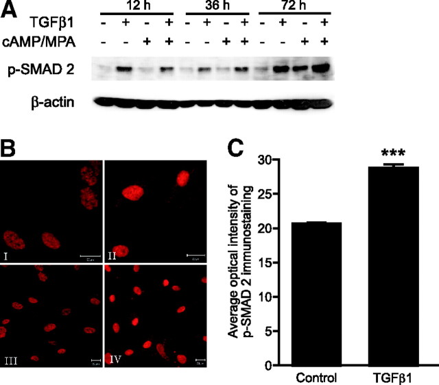
Fig. 6.
TGFβ1 Induces SMAD2 Phosphorylation in ESC, Both Decidualized and Nondifferentiated
A, Nuclear lysates from untreated primary ESCs or cultures decidualized in vitro for 12, 36, or 72 h, with or without TGFβ1 (10 ng/ml), were subjected to Western blotting analysis. TGFβ1 treatment induced protein expression of p-SMAD2 at each time point. B, ICF analysis of p-SMAD2 expression in control and TGFβ1-treated decidualized ESCs; I–IV, p-SMAD2 immunostaining; I, decidualized ESCs without treatment; II, decidualized ESCs plus TGFβ1 (10 ng/ml); III, decidualized ESCs without treatment, larger-view field; IV, decidualized ESCs plus TGFβ1 (10 ng/ml), larger-view field. C, Quantitative analysis of average optical intensity of p-SMAD2. Fifteen control and 15 TGFβ1-treated decidualized ESCs (10 ng/ml) were analyzed from each endometrial biopsy. All settings were identical for control and treated cells. TGFβ1 significantly up-regulated p-SMAD2 protein expression (P < 0.001); n = 5 endometrial biopsies. ***, P < 0.001.

