Fig. 4.
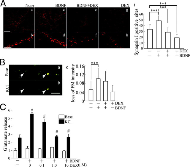
BDNF Failed to Increase the Number of Presynaptic Sites and the Release of Glutamate after DEX Pretreatment
A, The number of presynaptic sites was quantified after immunostaining with anti-synapsin I antibody. Top, Representative images from untreated cultures (none) (a) and cultures pretreated with BDNF (c), BDNF plus DEX (e), and DEX (g). Bar, 20 μm. Bottom, High-magnification images of dendrites from untreated cells (none) (b) and cells pretreated with BDNF (d), BDNF plus DEX (f), and DEX (h). Bar, 10 μm. i, Quantification of the number of synapsin I-positive presynaptic sites per dendritic shaft (50 μm). Data represent mean ± sd. None, n = 39; BDNF, n = 40; BDNF plus DEX, n = 46; DEX, n = 42. The n is the number of dendritic shafts selected randomly from cultured neurons for each experimental condition. BDNF increased the synapsin I-positive presynaptic number, and DEX treatment canceled the BDNF effect. Neurons at DIV16 were fixed after long maintenance in the presence or absence of BDNF or DEX, respectively. ***, P < 0.001, ANOVA. B, Effect of DEX on BDNF-potentiated presynaptic exocytotic activity. FM-43 images of before (a) and after (b) KCl (50 mm) stimulation. FM-dye signal revealed the presynaptic sites (arrowheads). The exocytotic activity in presynaptic sites was determined by elimination of the fluorescence. c, Quantification of reduction of FM-dye fluorescence. BDNF enhanced the elimination of FM-dye fluorescence. DEX had no influence. Data represent mean ± sd. The n (= 30) is monitored fluorescence dots in neurons for each experimental condition. The presynaptic sites of DIV7 neurons were monitored. Bar, 5 μm. ***, P < 0.001, Kruskal-Wallis test and Mann-Whitney U test. C, Amount of glutamate released from hippocampal cultures. White bars indicate the basal release of glutamate without stimulation. Black bars show the glutamate release after KCl stimulation. The reduction caused by DEX (0.1–10 μm) in the BDNF-potentiated glutamate release was observed. The glutamate release in DIV7 cultures (5 d after BDNF application) was measured and indicated as relative release compared with basal release in control (without DEX and BDNF). Data represent mean ± sd (n = 4). The n indicates the number of wells for each experimental condition on a plate. *, P < 0.05 (control vs. BDNF); #, P < 0.05 (BDNF vs. BDNF plus DEX), Kruskal-Wallis test and Mann-Whitney U test.

