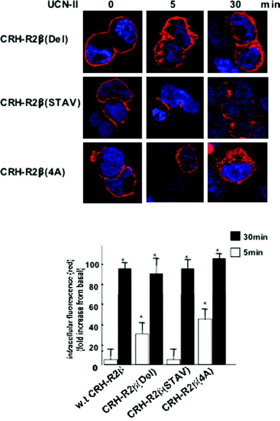
Fig. 12.

Role of CRH-R2β C-Terminal Amino Acid Cassette TAAV on Receptor Endocytosis
Cells were stimulated with 100 nm UCN-II for various time intervals, and CRH-R2β internalization was monitored by indirect confocal microscopy as described in Materials and Methods. Cell nuclei were stained with the DNA-specific dye DAPI (blue). Identical results were obtained from four independent experiments, and at least 20 cells were examined in each experiment. Inset, Fluorescence intensity of cytoplasmic CRH-R2β distribution. The sum of fluorescence intensity of cytoplasmic (distance 4–18 μm) fluorescence was measured by the ImageJ software. Results are expressed as the mean ± sem of three estimations from 20 individual cells. *, P < 0.05 compared with basal (unstimulated).
