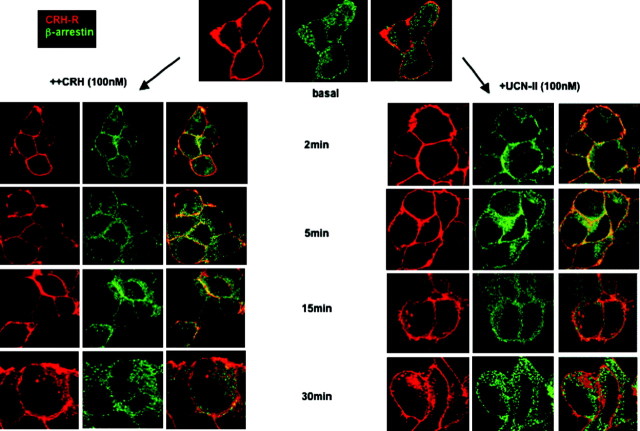
Fig. 3.
CRH-R2β and β-Arrestin Subcellular Distribution after CRH or UCN-II Treatment: Visualization by Fluorescent Confocal Microscopy
HEK293 cells expressing CRH-R2β were stimulated with either CRH or UCN-II (100 nm) for 2–30 min. CRH-R2β and β-arrestin distribution was monitored over the ensuing time period by indirect double immunofluorescence using specific primary antibodies and Alexa Fluor 594 secondary antibodies for CRH-R2β (red) and Alexa Fluor 488 secondary antibody for β-arrestin (green). Identical results were obtained from four independent experiments.

