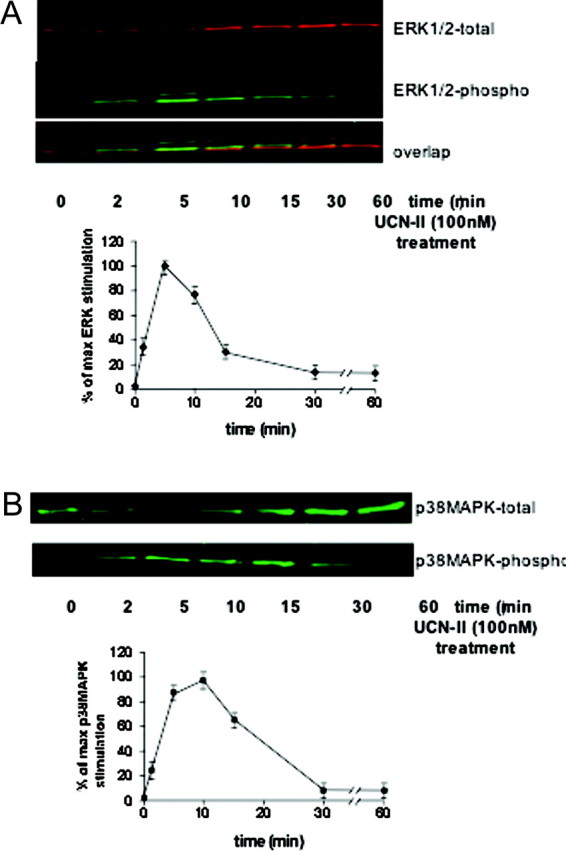
Fig. 8.

Time-Dependent ERK1/2 (A) and p38 MAPK (B) Activation by UCN-II in 293-R2β Cells
Top panels are representative Western blots of cells stimulated with UCN-II (100 nm) for various time points (2–60 min). After cell lysis and centrifugation, supernatants were subjected to SDS-PAGE and immunoblotted with antibodies for phospho-ERK1/2 and total ERK1/2 to determine the phosphorylated/activated ERK1/2 and secondary antibodies conjugated to IRDye800 and Alexa Fluor 680 (near-IR fluorophore dyes) as described in Materials and Methods. Alternatively, samples were immunoblotted with antibody for phospho-p38MAPK and total p38MAPK. Data represent the mean ± sem of three estimations from three independent experiments.
