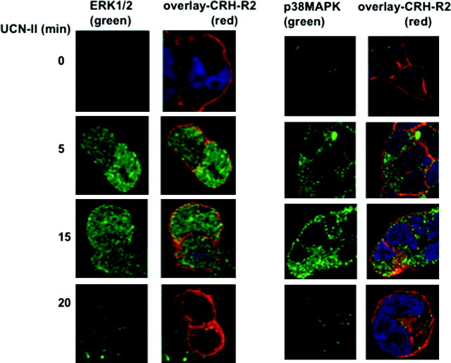
Fig. 9.
CRH-R2β and Phospho-ERK1/2 (A) or Phospho-p38MAPK (B) Subcellular Distribution Induced by UCN-II in 293-R2β Cells: Visualization by Confocal Microscopy
The 293-R2β cells were stimulated with or without UCN-II (100 nm) for various time intervals (5–20 min). CRH-R2β and phospho-ERK1/2 or phospho-p38MAPK distribution was monitored over the ensuing time period by indirect double immunofluorescence using specific primary antibodies for CRH-R2β and Alexa Fluor 594 secondary antibody (red) and Alexa Fluor 488 secondary antibody for phospho-ERK1/2 or p38MAPK (green) as described in Materials and Methods. Cell nuclei were stained with the DNA-specific dye DAPI (blue). Identical results were obtained from four independent experiments, and at least 20 cells were examined in each experiment. Scale bar, 20 μm.

