
Fig. 6.
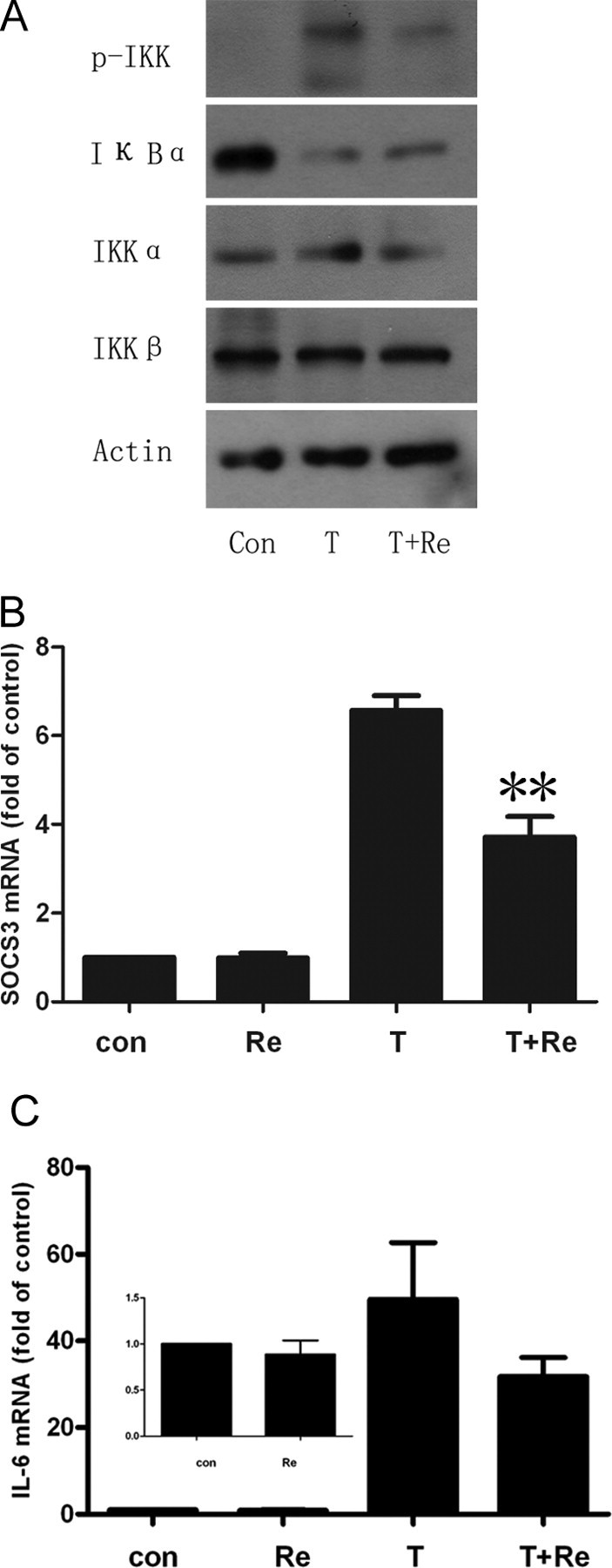
Inhibition of IKK/NF-κB Signaling Pathway by Re
A, Cells were treated with Re at 10 μm for 24 h and then with TNF-α at 1 nm for 30 min. The cell lysates were resolved by SDS-PAGE and analyzed using antibodies against total and phosphorylated IKKα/β and IκBα. Representative blots are shown from three independent experiments. B and C, Real-time PCR for SOCS-3 and IL-6 expression as described in Materials and Methods. 3T3-L1 adipocytes were incubated with 10 μm Re and with or without 10 ng/ml TNF-α for 24 h. SOCS-3 is significantly reduced and IL-6 remarkably decreased. **, P < 0.01 compared with TNF-α-treated group.