Fig. 2.

SF1 Is Phosphorylated by CDKs on S203
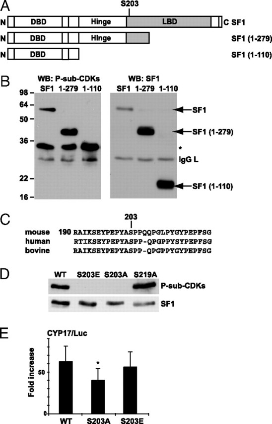
A, Schematic representation of full-length SF1 and the deletion constructs of SF1 [SF1(1–279), SF1(1–110)] used in this study. The numbering corresponds to the aa sequence. B, COS-1 cells were transfected with Flag-pCMV2/SF1 (SF1), Flag-pCMV2/SF1(1–110) (1–110), and Flag-pCMV2/SF1(1–279) (1–279), and SF1 was immunoprecipitated with the SF1 antibody and subjected to Western blot analyses using the anti-P-S-sub-CDKs antibody and subsequently the SF1 antibody. *, Unknown reactive protein. C, Sequence alignment of human, mouse, and bovine SF1. D, COS-1 cells were transfected with Flag-pCMV2/SF1 (WT) and mutants Flag-pCMV2/SF1(S203A) (S203A), Flag-pCMV2/SF1(S203E) (S203E), or Flag-pCMV2/SF1(S219A) (S219A). SF1 was immunoprecipitated with Flag antibodies, and Western blot analyses were performed using the anti-P-sub-CDKs antibody (upper panel) followed by incubation with SF1 antibodies (lower panel). E, COS-1 cells were transfected with CYP17/Luc (1 μg) together with Flag-pCMV2/SF1 WT, S203A, or S203E (100 ng). The luciferase activity is shown as fold increase and relative to β-gal activity (n = 7–10; *, P < 0.05 as determined by ANOVA, Bonferoni test). DBD, DNA-binding domain; WB, Western blot.
