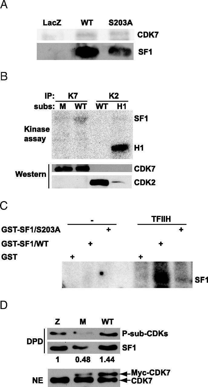
Fig. 4.

SF1 Coimmunoprecipitates with CDK7 and Is Phosphorylated by CDK7 and TFIIH
A, COS-1 cells were transfected with Flag-pCMV2/SF1 WT or S203A, or the control vector pCMV5/LacZ and immunoprecipitated with the Flag antibody. Eluates were run on a 10% SDS-PAGE and subjected to Western blot analyses using CDK7 (upper panel) and SF1 (lower panel) antibodies. B, CDK7 and CDK2 were immunoprecipitated from H295R cells and incubated with different substrates (subs), GST-SF1(179–431) (WT), GST-SF1(179–431/S203A) (M), or histone H1 (H1) for kinase assays. Membranes were subjected to autoradiography (O/N exposure) followed by Western blot analyses with anti-CDK7 and anti-CDK2. C, GST, GST-SF1(179–431), and GST-SF1(179–431/S203A) were incubated in the absence or presence of TFIIH isolated from HeLa cells. Gels were subjected to autoradiography (1–2 h exposure). D, H295R cells were transfected with control vector pCMV5/LacZ, Myc-CDK7 WT, or the Myc-CDK7-K41R (5 μg). SF1 was then isolated by DPD, and Western blotting was performed with anti-P-Sub-CDK (upper panel) and SF1 (middle panel) antibodies. Nuclear extracts (20 μg) were also subjected to Western blotting with anti-CDK7 (lower panel). Densitometric analyses were performed and are shown as ratios between phosphorylated SF1 and total SF1. The value from control cells was set to 1. IP, Immunoprecipitation; NE, nuclear extract.
