. 2017 Apr 11;6:e16059. doi: 10.7554/eLife.16059

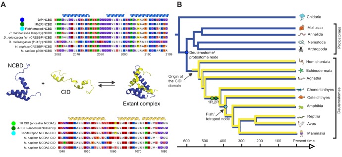
Figure 2. Reconstruction of the evolution of the interacting NCBD and CID domains.

(A) Sequence alignments of extant and reconstructed ancient NCBD (top) and CID domains (bottom). The positions of helices are according to the NMR structure of the complex between extant CREBBP NCBD (blue) and NCOA3 CID (yellow). Free NCBD (protein data base code 2KKJ) and the CID/NCBD complex (1KBH) are NMR structures, whereas the picture of free CID is a hypothetical modified structure made from the NCOA1 CID/NCBD complex (2C52). The first residue in the NCBD alignment is referred to as position 2062 in the text and the first residue in the CID alignment as 1040. The color coding of the sequences reflects similarities in chemical properties of the amino acid side chains and is a guide for the eye to see patterns of conservation. (B) Schematic tree of life with selected animal groups depicting the evolution of the NCBD domain (blue) in both protostomes and deuterostomes and the CID domain (yellow) in the deuterostome lineage only. See Figure 2—figure supplements 14 for detailed alignments and trees.

DOI: http://dx.doi.org/10.7554/eLife.16059.004

Figure 2—source data 1. Probabilities of resurrected amino acid residues at the respective position (2062–2109) in the NCBD domain.
elife-16059-fig2-data1.docx (125.3KB, docx)
DOI: 10.7554/eLife.16059.005
Figure 2—source data 2. Probabilities of resurrected amino acid residues at the respective position (1040–1081) in the CID domain.
The gap in Figure 2—figure supplement 4 created by one of the Takifugu rubripes sequences was removed in this table to make it easier to understand the numbering.
elife-16059-fig2-data2.docx (125.5KB, docx)
DOI: 10.7554/eLife.16059.006

Figure 2.

Figure 2—figure supplement 1. Sequence alignment of NCBD domains of CREBBP/p300 used in the phylogenetic reconstruction.
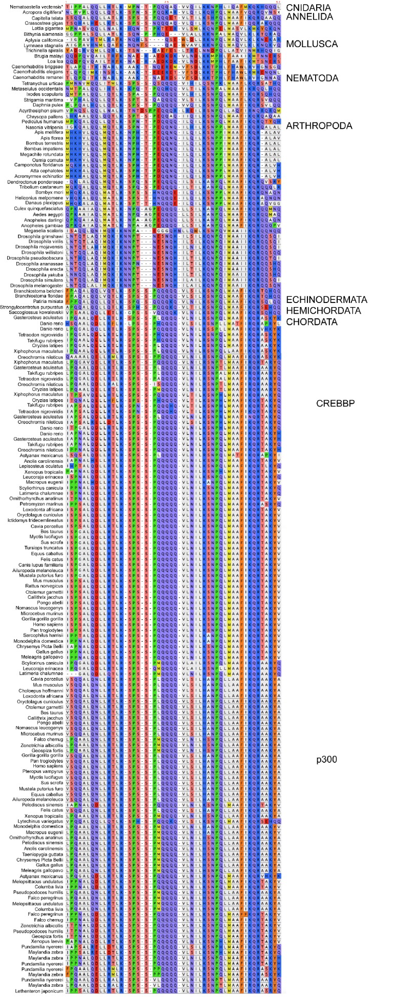
Figure 2—figure supplement 1.

The whole CREBBP/p300 gene alignment was used to create the phylogenetic tree and then with the use of that tree the NCBD domain could be resurrected. The categories to the right should only be seen as guidelines to which group the individual species sequences belongs. Amino acids are colored based on chemical properties of the side chain according to eBioX standard. The tree in Figure 2—figure supplement 3 has the correct grouping information.
Figure 2—figure supplement 2. Sequence alignment of the CID domains of NCOA1-3 used in the phylogenetic reconstruction.
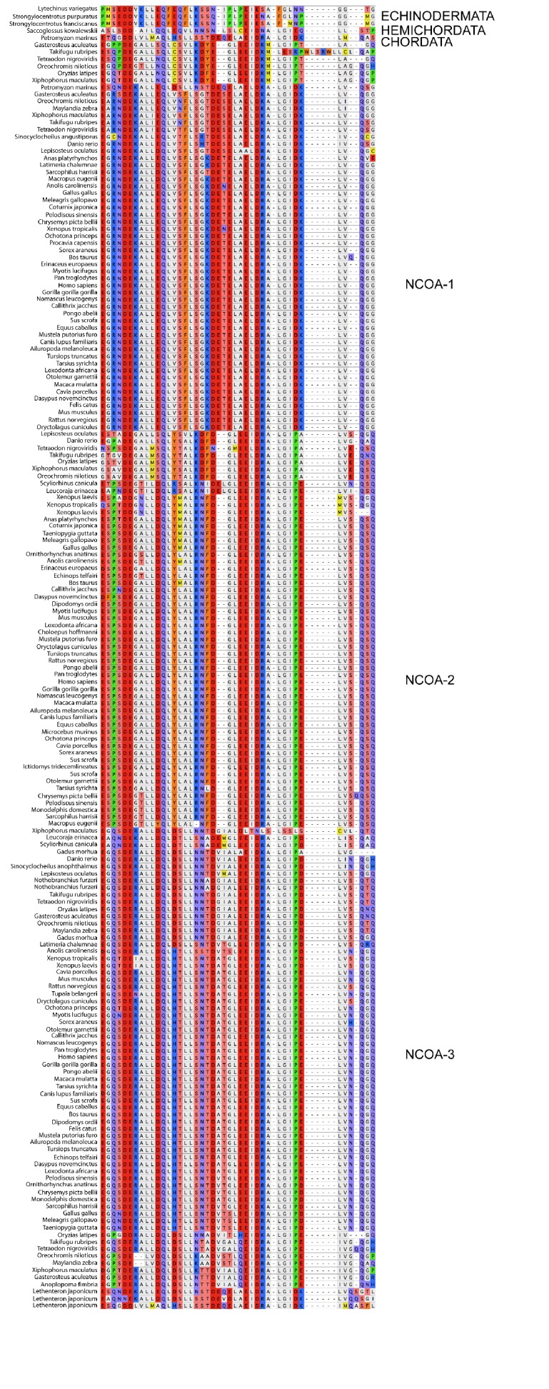
Figure 2—figure supplement 2.

The whole NCOA1-3 gene alignment was used to create the phylogenetic tree and then with the use of that tree the CID domain could be resurrected. The categories to the right should only be seen as guidelines to which group the individual species sequences belongs. Amino acids are colored based on chemical properties of the side chain according to eBioX standard. The tree in Figure 2—figure supplement 4 has the correct grouping information.
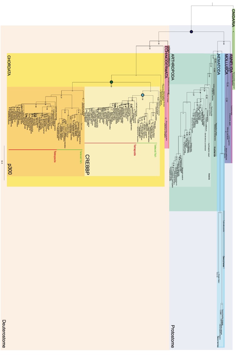
Figure 2—figure supplement 3. Phylogenetic tree of CREBBP/p300 proteins that contain the NCBD domain.

Figure 2—figure supplement 3.

The tree follows what is generally accepted regarding species evolution and whole genome duplications. Cnidarian CREBBP/p300 contains the NCBD domain. Since these species are distantly related to the other animals in the tree Cnidarian CREBBP/p300 proteins are used as outgroup in the analysis. After the divergence of Cnidaria from other metazoans, deuterostomes and protostomes diverged from each other. The node between the deuterostomes and protostomes (marked with a dark blue circle) is the oldest time point that we resurrect. Protostomes contain groups such as insects and molluscs, while deuterostomes contain all vertebrates. At the beginning of vertebrate evolution two whole genome duplications occurred. Thus, at this point all vertebrate genes were duplicated twice resulting in four copies, but many were rapidly lost. For CREBBP/p300, two copies remained, namely CREBBP and p300. The node corresponding to the ancestral CREBBP/p300 protein is marked with a green circle and referred to as 1R/2R in the text, since we cannot distinguish these events for CREBBP/p300. The most recent node that we resurrect is the separation of Teleost fish CREBBP from Tetrapod CREBBP (marked with a light blue circle). A third round of whole genome duplications later occurred in the fish lineage (not indicated) resulting in the two variants CREBBP1 and CREBBP2. The indicated resurrected nodes correspond to the same nodes as in the simplified Figure 2 in the main text.
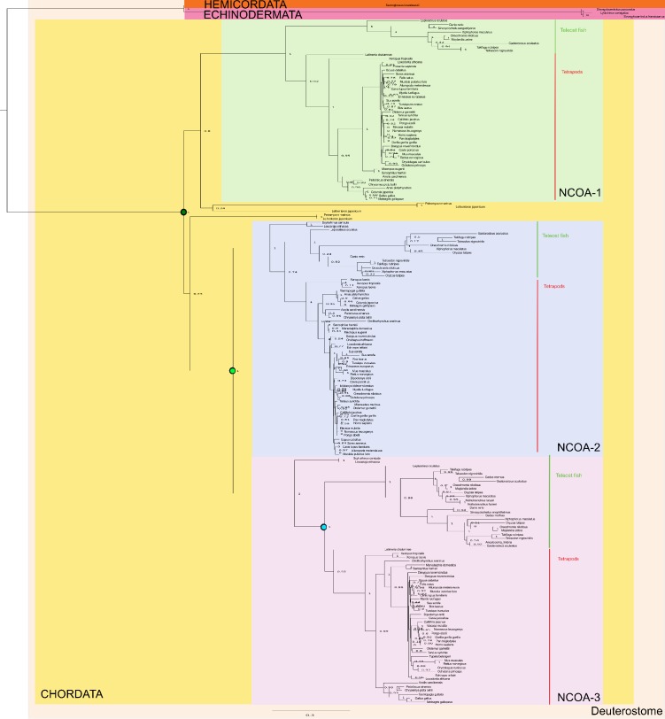
Figure 2—figure supplement 4. Phylogenetic tree of NCOA1-3 proteins that contain the CID domain.

Figure 2—figure supplement 4.

The tree follows what is generally accepted regarding species evolution and whole genome duplications. The CID domain could be identified in Hemicordata and Echinodermata NCOA proteins, and since these species are more distantly related to the other animals in the tree they were used as outgroups in the analysis. After the divergence of these groups from the rest of the deuterostomes, the two vertebrate-specific whole genome duplications occurred. Our analysis suggests that the ancestral NCOA gene was first split into NCOA-1 and the ancestor of NCOA2 and 3, which were subsequently split into NCOA2 and NCOA3 in the second whole genome duplication. (The second copy of NCOA1 was lost from the genomes) The nodes are marked with a dark (1R) and light green circle (2R), respectively. The most recent node that we resurrected is the separation of Teleost fish NCOA3 from Tetrapod NCOA3 (marked with a light blue circle). A third round of whole genome duplications occurred later in the fish lineage (not marked) but the resulting NCOA proteins were not retained in the genome. The indicated resurrected nodes correspond to the same nodes as in the simplified Figure 2 in the main text.