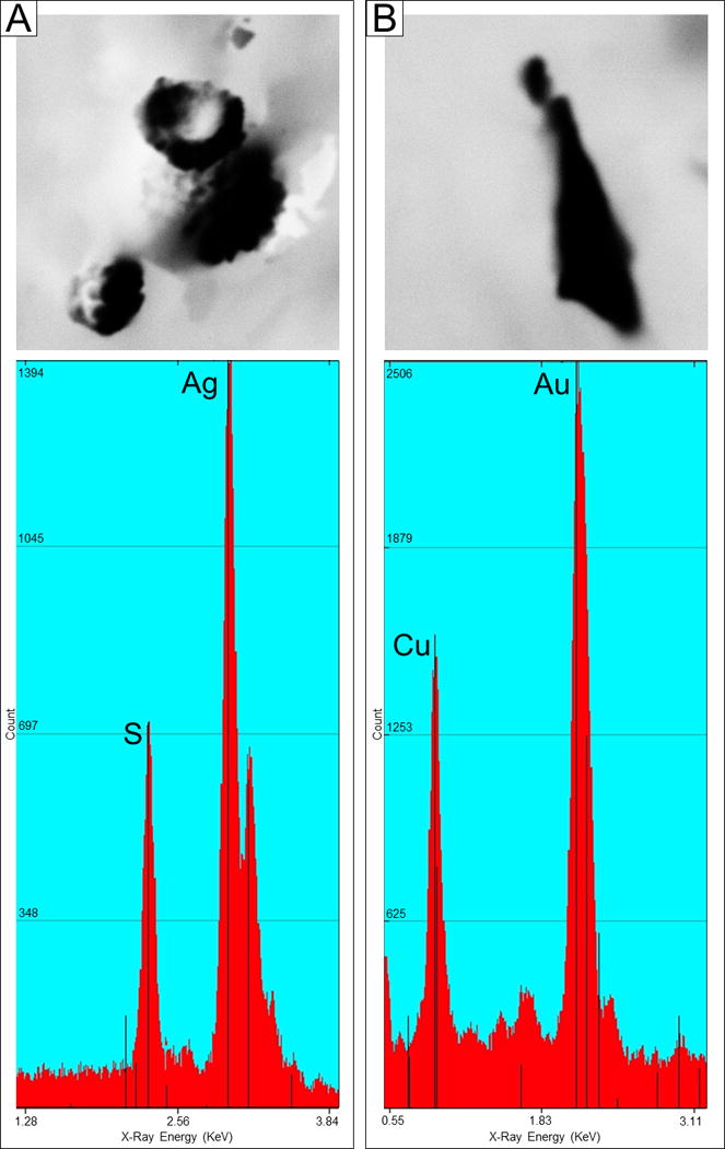
Figure 2. Backscatter Electron Imaging and Energy Dispersive X-Ray Analysis.

a. Backscattered electron imaging (BEI), 2700×, irregularly shaped AgS particles; Spectrum showing peaks for Ag and S
b. BEI, 7000×, irregularly shaped AuCu particle; Spectrum showing peaks for Au and Cu
