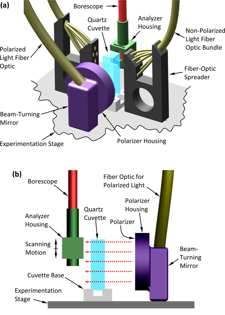
Figure 2.
Schematic illustration of the experimentation stage of the scanning cryomacroscope incorporating polarized-light means: (a) isometric view including all illumination components, (b) side view showing polarized-light components only, where the red-dashed arrows represent the direction of polarized light illumination.

