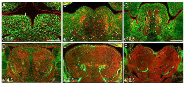
Figure 2. NCC contribution to the glossal mesenchyme.
(A–F) Frontal sections through the developing tongue of ROSAmT/mG;Wnt1-Cre embryos between e10.5 and e18.5. NCCs are green and non-NCCs are red. (A) At e10.5 the lateral lingual swellings (white arrows) are primarily composed of NCCs. (B–C) Between e11.5 and e12.5 non-NCC-derived presumptive myoblasts (red) migrate into the tongue anlage (dotted white lines). (D–F) Between e14.5–e18.5 the non-NCC-derived muscle fibers organize and populate the majority of the tongue. Scale bars = 250um.

