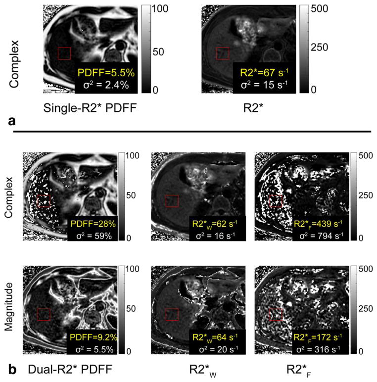
FIGURE 9.
Dual-R2* fitting to in vivo data has artifacts and high standard deviations. All reconstructions shown were performed on the same slice at 1.5T. The red ROI is colocalized with the spectroscopy voxel (spectroscopy FF = 6.3%). (a) Single-R2* correction results in PDFF = 5.5% and R2* = 67s−1. The maps have no noticeable artifacts in the liver. (b) Dual-R2*, for both complex and magnitude fits, results in artifacts and high standard deviations in PDFF, R2*W, and R2*F maps. These measurements were deemed unreliable and further statistical analysis was not performed on the dual-R2* reconstructions.

