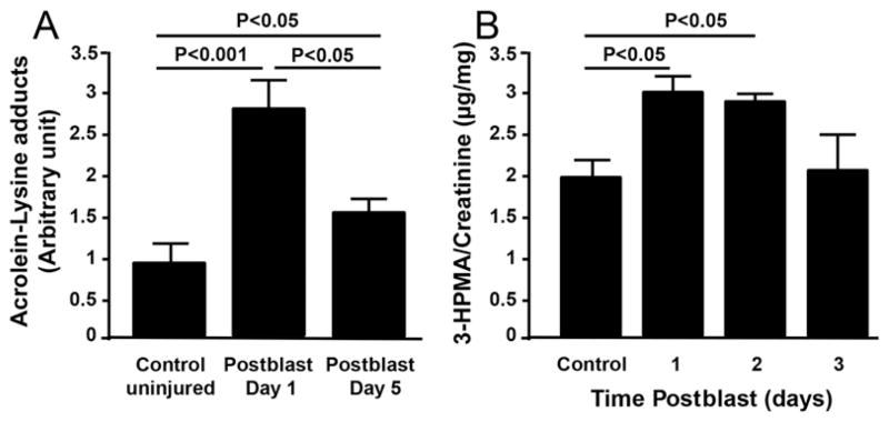
FIG. 7.
Biochemical markers for tissue and cellular stress. A: Acrolein-lysine adducts in the brain tissue at 1 (Day 1) and 5 (Day 5) days postblast were detected via dot blot analysis. The acrolein-lysine level of both Day 1 and Day 5 are significantly elevated when compared with levels in preblast controls (p < 0.001 and p < 0.05, respectively, Tukey-Kramer test; n = 5 rats/group). In addition, the acrolein-lysine level of Day 1 is significantly higher than that of Day 5 (p < 0.05, Tukey-Kramer test; n = 5 rats/group). B: Urine 3-HPMA detected by LC-MS/MS relative to urine creatinine content. Urine was collected for 3 days postblast, indicating significant elevation of 3-HPMA in the first 2 days following exposure as compared with preexposure levels (p < 0.05, Tukey-Kramer test; n = 5 rats).

