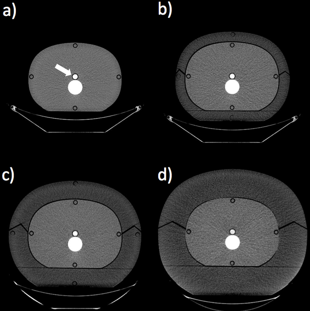
Figure 1.

Axial CT images showing the four phantom size configurations. The white arrow shows the location of the contrast vial, immediately anterior to the spine insert (Window Width = 400; Window Level = 40). a) Base phantom composed of soft-tissue-equivalent plastic, outer diameter = 220 × 300 mm. b) Base phantom with a 33-mm-thick fat ring composed of adipose-equivalent plastic, outer diameter = 285 × 365 mm. c) Base phantom with a 60-mm-thick fat ring, outer diameter = 340 × 420 mm. d) Base phantom with a 90-mm-thick fat ring, outer diameter = 400 × 480 mm.
