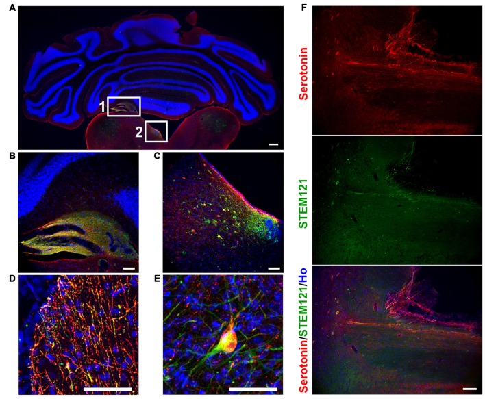
Figure 2.
Human iPSCs-derived serotonergic neurons migrate and project to the host’s cerebellum, hindbrain and spinal cord in vivo. (A–C) After 3 months of transplantation, human serotonergic neurons formed chimera with the host’s brain tissues around the 4th brain ventricle. (B) is the detailed image for inset 1 in (A); (C) is the detailed image for inset 2 in (A). (D) Serotonin+ human fibers were observed in the host’s cerebellum. (E) Serotonin+ human cell bodies were observed in the host’s ventral hindbrain. (F) Serotonin+ human fibers were observed in the host’s spinal cord. Scale bar: (A) 200 μm; (B–F), 50 μm; Red: Serotonin+ cells; Green: STEM121+ cells (human cells); Blue: Hoechst staining (Ho).

