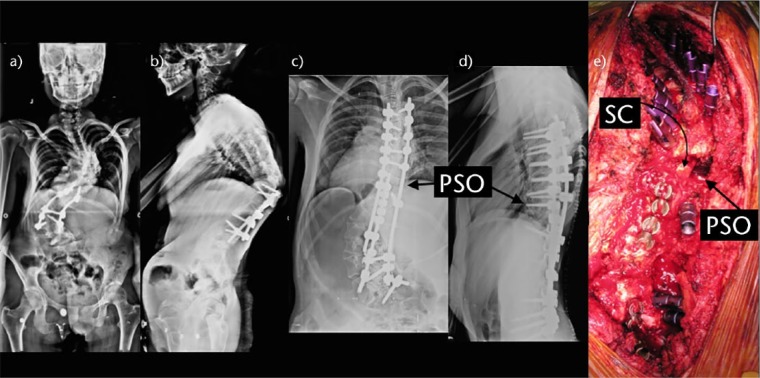
Fig. 4.
Pedicle subtraction osteotomy in revision scoliosis surgery. a, b) Anteroposterior (AP) and lateral views of a 21-year-old female patient who had undergone a previously unsuccessful fusion operation. She presented with both a severe scoliosis and also decompensated kyphosis. The dotted white line represents the planned osteotomy. c, d) AP and lateral views after surgery. There was a significant correction of both coronal and sagittal balance. The osteotomy sites are shown with black arrows. e) Intra-operative view of an asymmetric T10 pedicle subtraction osteotomy and instrumentation. Spinal cord and osteotomy site are shown with black arrows. SC, spinal cord; PSO, pedicle subtraction osteotomy.

