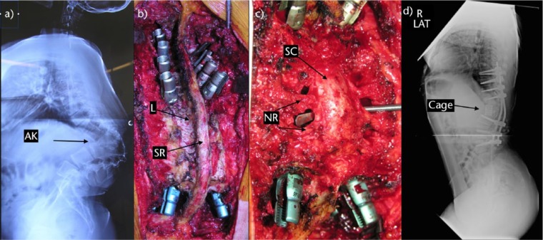
Fig. 8.
a) A 33-year-old woman with congenital kyphosis. b) Intra-operative view of the kyphotic region. c) Intra-operative view of three levels. Vertebral column resection including T11, T12 and L1. d) Post-operative lateral view. AK, apex of kyphosis; L, lamina; SP, spinous process; NR, nerve root; SC, spinal cord.

