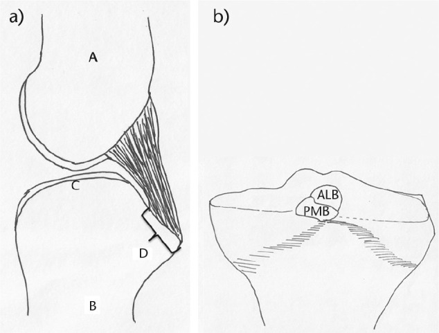
Fig. 2.

The two bundles of the posterior cruciate ligament (PCL) are very compact and difficult to separate at their tibial origin. a) Sagittal view of the tibial origin of the PCL. A, femur; B, tibia; C, tibial articular surface; D, origin of PCL. b) Coronal view of the posterior aspect of the proximal tibia showing the origin of the anterolateral bundle (ALB) and posteromedial bundle (PMB).
