
Fig. (4).
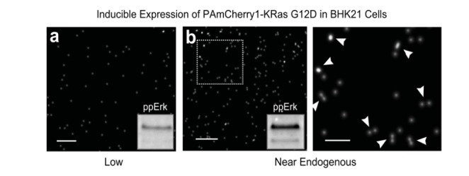
Single-molecule superresolution imaging of Ras dimers in cells. BHK21 cells stably expressing PAmCherry1-KRas G12D (an activated mutant of KRas that constitutively binds GTP) under doxycycline (Dox) regulation was treated with 1 or 2 ng/mL Dox for 48-72 hours before being fixed and imaged with photoactivated localization microscopy (PALM). Images were acquired under total internal reflection (TIR) illumination conditions to limit the excitation volume to the basal membrane of the cells. Each dot in the PALM images represents one putative PAmCherry1-KRas G12D molecule. (a) At 1 ng/mL Dox, PAmCherry1-KRas G12D is expressed at a level much lower than that of endogenous KRas and appears monomeric, when the level of phosphorylated Erk (ppErk) is also low as shown in the inset, indicating little activation of the Raf-MAPK signaling pathway; (b) At 2 ng/mL Dox, PAmCherry1-KRas G12D is expressed at a level similar to that of endogenous KRas, and dimers (and occasional higher order multimers) of PAmCherry1-KRas G12D could now be observed. Image on the right is the zoomed view of the boxed area in the image on the left. White arrows indicate putative KRas dimers. Under this condition, ppErk level is significantly higher, indicative of an activated Raf-MAPK pathway. Scale bars, 250 nm in (a) and (b, left), and 100 nm in the zoomed view (b, right).