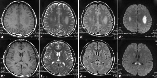
Figure 1.
Brain magnetic resonance imagings of a male patient with X-linked Charcot-Marie-Tooth type 1 (patient 13) revealed abnormal signals in the bilateral centrum semiovale (a–d) and splenium of the corpus callosum (e–h). (a and e): T1 weighted; (b and f): T2 weighted; (c and d): Fluid attenuated inversion recovery; and (d and h): Diffusion-weighted magnetic resonance imaging.

