Figure 1.

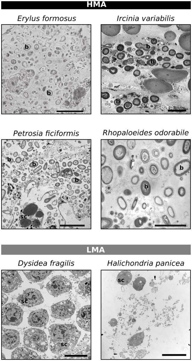
Classification of the HMA-LMA status of sponges based on transmission electron microscopy. Scale bars represent 5 μm, but vary in length. b, bacteria; sc, sponge cell.

Classification of the HMA-LMA status of sponges based on transmission electron microscopy. Scale bars represent 5 μm, but vary in length. b, bacteria; sc, sponge cell.