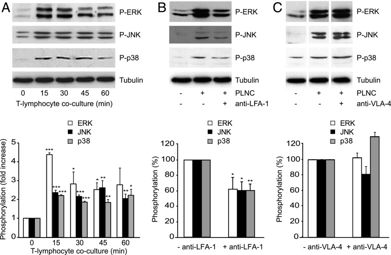
FIGURE 1.
Endothelial MAPK activation in response to lymphocyte adhesion. (A) All three MAPKs were activated in GPNT ECs cocultured with Con A–activated, nonmigratory rat PLNCs. Shown are representative immunoblots of MAPKs phosphorylation alongside tubulin loading controls and normalized densitometric quantification of three independent experiments. (B and C) MAPK activation in GPNT in 30 min cocultures was reduced when PLNCs were preincubated with function-blocking anti–LFA-1 Abs but not an anti–VLA-4 blocking Ab. Shown are representative blots and densitometric quantification of three independent experiments. Control phosphorylation levels (in response to PLNC adhesion without adhesion molecule neutralization) were set to 100%. Data were compared with the corresponding time 0 controls and significant differences are indicated. In (C), white separation lines indicate where lanes from the same blots were joined. *p < 0.05, **p < 0.01, ***p < 0.001.

