FIGURE 5.
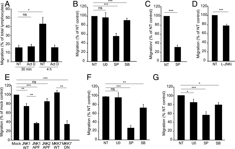
Endothelial JNK regulates lymphocyte TEM. (A) GPNT monolayers were pretreated or not with actinomycin D (Act D, 5 μg/ml) and subsequent TEM of Th1 lymphocytes (PAS, see also Supplemental Fig. 3A) measured after 30 min. Whereas Act D did not affect 30 min TEM rates, it inhibited all subsequent TEM events (measured up until 4 h). (B) TEM assay as in (A) with the exception that GPNT monolayers were left untreated (NT) or treated with 50 μM U0126 (U0), SP600125 (SP) or SB202190 (SB) for 1 h prior to a 30 min TEM assay. Due to the high washout rate of U0126 from GPNT cells (see Supplemental Fig. 2B), TEM experiments were also conducted with U0126 present throughout. However, even under these conditions TEM was not inhibited (data not shown). (C) TEM assay as in (B) except that primary rat brain MVEC were either left untreated (NT) or pretreated with 50 μM SP600125 for 1 h prior to addition of Ag-specific T lymphocytes. (D) TEM assay as in (B) with the exception that GPNT were either left untreated (NT) or treated with 1 μM L-JNKi for 1 h prior to the addition of T lymphocytes. (E) TEM assay as in (B) except that GPNT cells were transfected with wild-type (WT) or dominant-negative (DN) JNK1, JNK2, or MKK7 48 h before TEM and adhesion were analyzed. (F and G) TEM assay as in (B) except that TEM of human CD4+ cells across hCMEC/D3 (F) or human dermal MVEC (G) was measured following EC pretreatment with 50 μM U0126 (U0), SP600125 (SP), or SB202190 (SB) for 1 h. *p < 0.05, **p < 0.01, ***p < 0.001.

