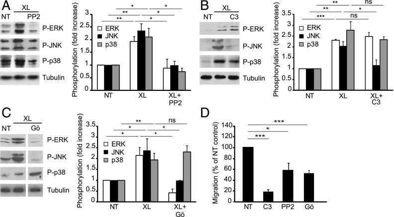
FIGURE 6.
Role of Src, Rho GTPase, and PKC in ICAM-1–mediated MAPK activation and lymphocyte TEM. Postconfluent, serum-starved GPNT cells were either left untreated (NT) or pretreated with 10 μM PP2 for 30 min (A), 10 μg/ml C3 transferase for 12 h (B), or 20 μM Gö6983 (Gö) for 30 min (C). In (C) white separation lines indicate where lanes from the same blots were joined. Where indicated EC monolayers were subjected to ICAM-1 cross-linking (XL) for 10 min. MAPK phosphorylation was then analyzed and quantified as described for Fig. 1. Results similar to those shown with PP2 were also found with 10 μM SU6656 (data not shown). MAPK levels were not significantly affected by any of the pretreatments (Supplemental Fig. 1C). (D) GPNT EC monolayers were pretreated with 10 μM PP2 for 1 h, 10 μg/ml C3 transferase for 16 h, or 20 μM Gö6983 (Gö) for 1 h prior to analysis of lymphocyte TEM. *p < 0.05, **p < 0.01, ***p < 0.001.

