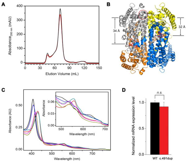
Figure 2.
(A) Superdex-200 elution profile of the wild-type enzyme (black trace) and Met108Ile (red trace). (B) Structural representation of the distance between Met108 and the heme center (34 Å) in the same subunit and adjunct unit (32 Å). The coordinates were taken from 5TIA.PDB [21]. (C) The optical spectra of Met108Ile at the ferric (black trace), ferrous (red) oxidation state, ferric-CN complex (magenta), and in the presence of L-Trp with the ferric (blue), ferrous (cyan), and ferric-CN complex (brown), respectively. The inset is a zoomed-in view of the α/β band region. These spectra are indistinguishable from those of the wild-type enzyme. (D) The mRNA expression level of c.491dup and the native enzyme in HeLa cells as quantitated by qPCR.

