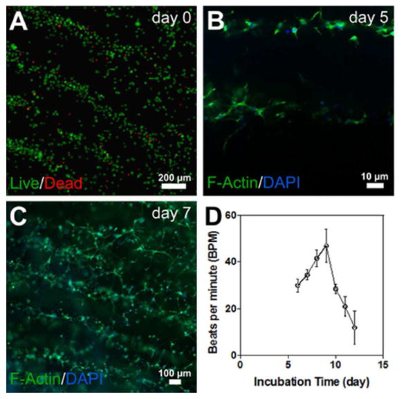
Figure 5.

A) Viability assay on day 0 reveals nearly 90% live cells; B) F-actin/DAPI staining of cells after 5 days, when the encapsulated patterned cardiac cells demonstrated spreading; C) F-actin/DAPI staining of cells after 7 days, when the encapsulated patterned cardiac cells started forming interconnected cellular networks; D) Spontaneous beating rates of 3D cardiac tissues over time.
