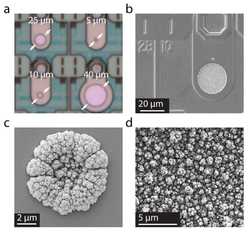
Figure 2.
(a) Microscopic picture of the platinum electrodes with different diameters. The white arrows denote the active area of the working electrodes. (b, c) Scanning electron micrographs of a 25 μm-diameter electrode (b) and 5 μm-diameter electrode (c) modified by Pt black. (d) Magnified cauliflower-like structure of the Pt black surface. (b, c, d) Picture: D. Mathys, Centre of Microscopy (ZMB), University of Basel Klingelbergstrasse 50/70, CH-4056 Basel.

