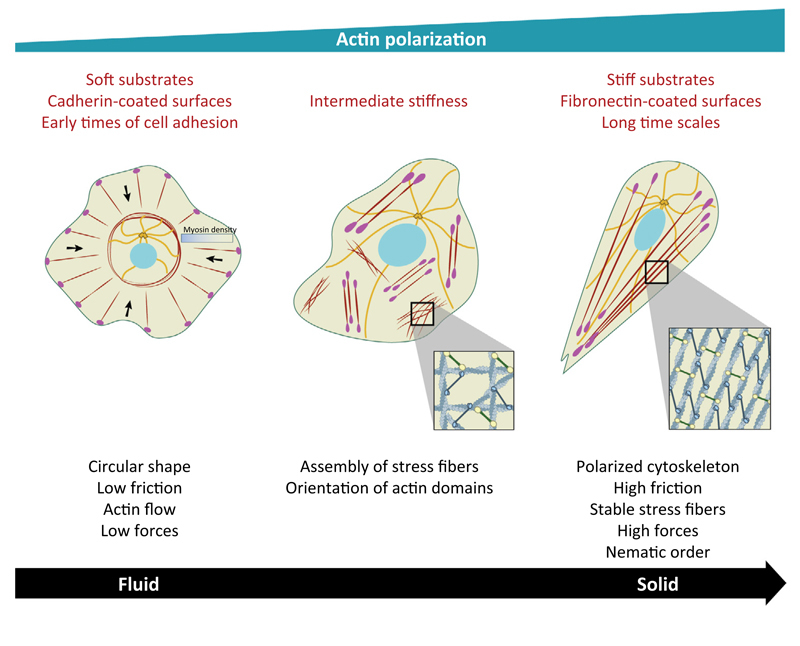
Figure 1. Single Cell Polarization by Biomechanical Cues.
(Left) Circular shape of a single cell under various conditions with the formation of noncontractile actin radial fibers at the edge and transverse arcs at the back (actin filaments are in dark red). Microtubules (MTs; orange) are confined in the central region. Light red dots represent focal adhesions (FAs) and black arrows represent the direction of the actin retrograde flow. (Middle) Appearance of ventral stress fibers that are organized in local domains on substrate of intermediate stiffness. The order parameter, S, that characterizes actin orientation is low. Note MTs reaching the edge of the cells. (Right) Actin polarization characterized by a large-scale alignment of actin filaments on stiff substrates, at long time scales and/or on fibronectin-coated surfaces (S = 1).

