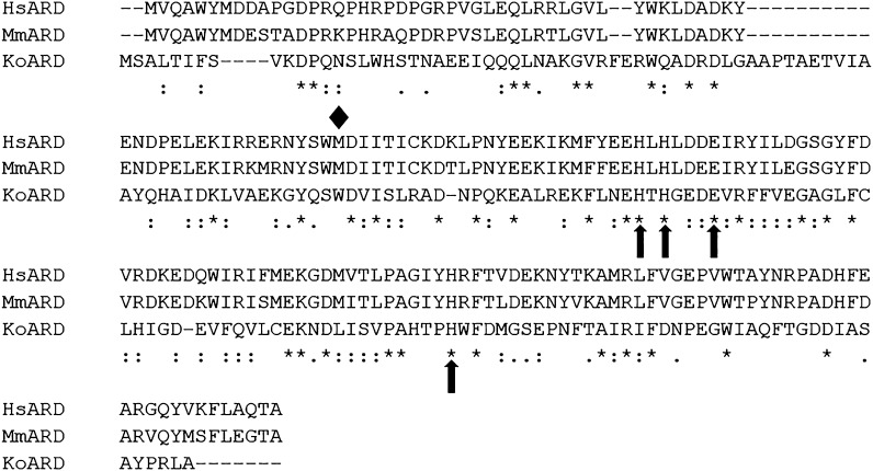
Fig. 3.
Sequence alignment of ARD homologs. The metal-binding residues are marked by arrows and the diamond represents the first methionine residue of N-terminal truncated HsARD denoted as SiPL which is implicated in replication of hepatitis C virus in non-permissive cell lines. Sequence alignment was performed using ClustalOmega.

