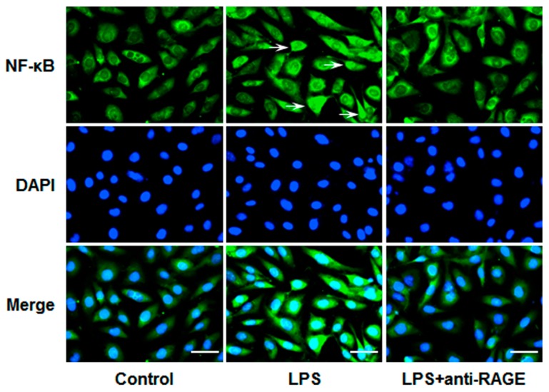
Figure 4.
HUVECs grown on Petri dishes were pretreated with anti-RAGE (10 μg/mL) for 1 h and followed by LPS (1 μg/mL) stimulation for 12 h. Then cells were processed for immunostaining with anti-NF-κB p65 antibody. Nuclei of cells were stained with DAPI (blue) and p65 was visualized by green fluorescence. The arrows show the nuclear localization of NF-κB. Scale bars, 50 μm. Results are representative images of three independent experiments.

