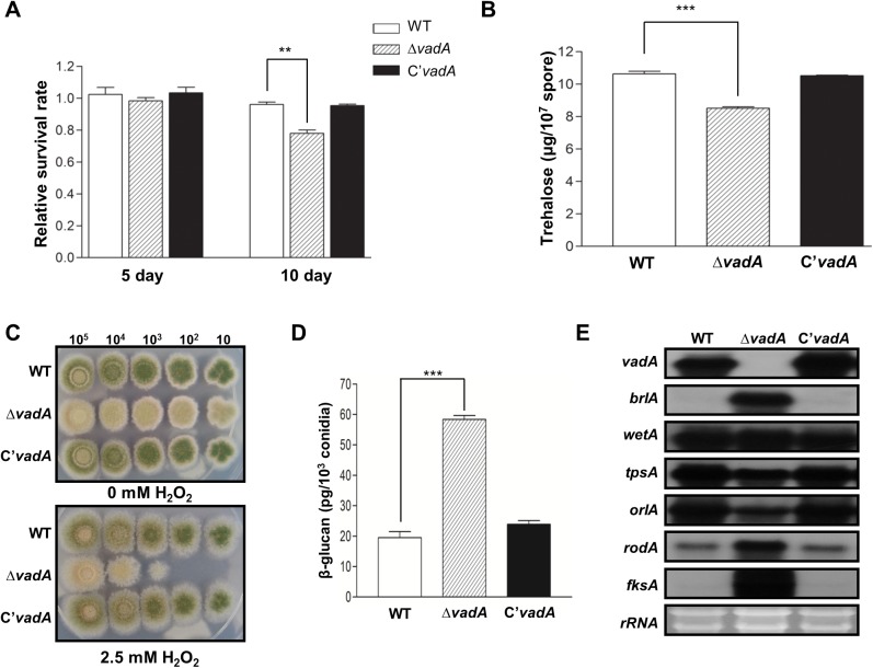
Fig 3. The role of vadA in conidia.
(A) Viability of the conidia of WT (FGSC4), ΔvadA (THS33.1), and C’ (THS34.1) strains grown at 37°C for 5 and 10 days (** P < 0.01). (B) Amount of trehalose per 107 conidia in the two-day-old conidia of WT (FGSC4), ΔvadA (THS33.1), and C’ (THS34.1) strains (measured in triplicate) (*** P < 0.001). (C) Tolerance of the conidia of WT (FGSC4), ΔvadA (THS33.1), and C’ (THS34.1) strains to H2O2 treatment. (D) Amount of β-glucan (pg) per 103 conidia in the two-day-old conidia of WT (FGSC4), ΔvadA (THS33.1), and C’ (THS34.1) strains (measured in triplicate). (E) Levels of vadA, brlA, wetA, tpsA, orlA, rodA, and fksA transcripts in the conidia of WT (FGSC4), ΔvadA (THS33.1), and C’ (THS34.1) strains. Equal loading of total RNA was confirmed by ethidium bromide staining of rRNA.

