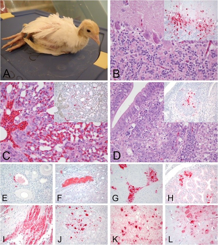
Fig 6. Clinical presentation, histopathology and immunohistochemical staining for avian influenza virus antigen in tissues of turkeys infected with the H7N8 HPAIV, 2days post infection.
All photomicrographs, magnification 40X; hematoxylin and eosin staining (B-D); immunohostchemistry (insets in B-D, and E-L), virus staining in red. A. Turkey with neurological signs. B. Cerebellum. Severe multifocal neuronal necrosis. Insert: Cerebellum, same area. Viral antigen staining in neurons, Purkinje cells, and glial cells. C. Lung. Moderate lymphoplasmacytic interstitial pneumonia. Inset: Lung, same area. Viral antigen staining in epithelium of air capillaries and mononuclear cells. D. Cloacal bursa. Lymphoid depletion with necrotic/apoptotic lymphocytes. Inset: Cloacal bursa, same area. Viral staining in macrophages in the medulla. E. Ovary. Viral staining in tegument/interstitial tissue and ova epithelium. F. Kidney. Viral staining in tubular epithelial cells. G. Adrenal gland. Viral staining in corticotrophic cells. H. Proventriculus. Viral antigen staining in epithelium of the proventricular gland. I. Heart. Viral staining in myocytes. J. Pancreas. Viral antigen staining in mononuclear cells. K and L. Cerebrum. Viral staining in neurons and glial cells.

