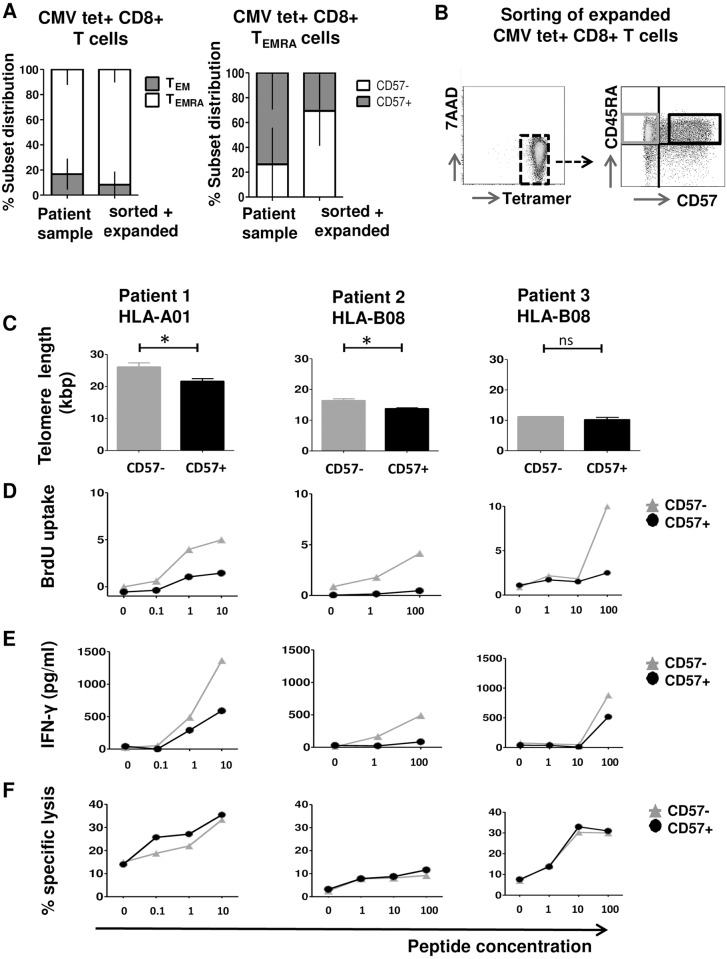
Fig 3. Phenotypic and functional characterization of CMV tetramer+ cells.
(A) CD8+ CMV tetramer+ T cells were FACS sorted from the peripheral blood of 3 patients after allogeneic SCT and in vitro expanded on autologous feeder cells. Depicted is the TEM and TEMRA subset distribution within CD8+ CMV HLA/tetramer+ T cells (left) and CD57+ distribution within CD8+ CMV HLA/tetramer+ TEMRA cells (right) in the peripheral blood compared to after in vitro expansion of FACS sorted CD8+ CMV tetramer+ T cells. Y-axis: % subset distribution within CD8+ CMV HLA/tetramer+ T cells and CD8+ CMV HLA/tetramer+ TEMRA cells. Error bars indicate standard deviation. (B) Sorting strategy for viable in vitro expanded CD8+ CMV HLA/tetramer+ CD8+ T cells for CD45RA and CD57 allowing functional analysis. (C) Absolute telomere length directly after sorting. (D) BrdU uptake 4 days after stimulation with CD14+ monocytes loaded with increasing concentrations of the relevant HLA/CMV peptide. (E) INF-γ release in the supernatant from the BrdU uptake assay. (F) Specific lysis of CFSE labelled PHA blasts loaded with increasing concentrations of the relevant HLA/CMV peptide. Significance was calculated using Mann-Whitney-U test. * indicates p<0.05, ** indicates p<0.01, ns indicates not significant.

