
Figure 5. Autophagy inhibitor 3-MA worsens theinjury of high glucose in Bim reduced cells by re-trigger apoptosis.
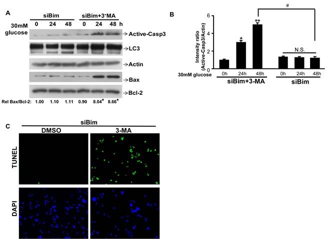
A. HK2 were transfected with siBim RNA for Two days, then the cells wereco-treated with 3-MA (or DMSO) and 30mM glucose for indicated times and lysed. Protein levels of active-Caspase3, LC3, Actin, BAX and BCL-2 were examined by immunoblotting. Quantification of the expressionratio of BAX/BCL-2 is shown with the ratio of 1.0 being assigned to siBim-0h cells. *P < 0.05; one-way ANOVA. B. Active-Caspase3/Actin immunoreactivityintensity was quantitated by densitometric analysis, and optical density values were expressed as a ratiobetween the Active-Caspase3 and Actin. Data shown are the mean ± s.e.m. n = 3. *P < 0.05, **P < 0.01, #P < 0.05, Student's t test.N.S. indicates not significant. C. HK2 were transfected with siBim RNA for Two days, then the cells wereco-treated with 3-MA (or DMSO) and 30mM glucose for 48h. Apoptosis was determined by TUNEL staining (green dots) and doubly stained with DAPI (blue dots).