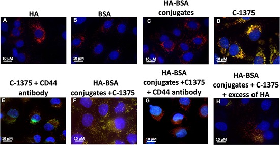
Figure 12.

Ovarian carcinoma SKOV3 cells were incubated with the following samples: (A) HA; (B) BSA; (C) HA-BSA conjugates; (D) C-1375; (E) C-1375 with blocking antibody against CD44; (F) HA-BSA loaded with C-1375; (G) HA-BSA loaded with C-1375 + CD44 antibody; and (H) HA-BSA loaded with C-1375 + free HA (molar ratio of free HA to conjugated HA was 100:1) for 1 h at 37°C. BSA-HA NPs loaded with C-1375 (10 μM) were added for 2 hr prior to fluorescence cell imaging (CD44 antibody (10 μg/ml) was added for 1 hr prior to BSA-HA conjugates). LysoTracker red DND99 (100 nM) was added for 1 hr prior to fluorescence cell imaging. The viable DNA dye Hoechst 33342 (2 mg/ml) was added immediately prior to microscopic analysis. Cells were then photographed using the Cell-Observer microscope at ×630 magnification.
